Article
More About Physical Therapy

X-ray vision is highly inconvenient. Think about it. Government agents and mafia criminals would try to force your allegiance and Christmas presents would never be a surprise. Good thing we don’t need superhero powers to see inside the human body. More than just exposing anatomy, diagnostic imaging provides medical professionals with key understanding into your health: something even Superman can’t do. There are three common imaging procedures we will explore below: X-rays, CT scans, and MRIs.
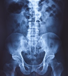
Radiographs (a.k.a. X-rays) utilize an electromagnetic beam to view our body structures. These structures are illuminated depending on how much X-ray they absorb. Bones absorb a lot of ray so they appear the whitest compared to air (think lungs) and soft tissues (organs, muscle). X-rays not only spot bone fractures but they can also expose cancer, lung disease, and other pathologies. The big fear with undergoing X-rays is radiation exposure. However the amount and intensity of radiation exposure is indescribably small compared to potentially harmful quantities. Most medical professionals agree that the benefits of X-rays always outweigh the risks.
CT scans (a.k.a. CAT scans) are a more detailed X-ray. CT scans involve a specialized X-ray machine shaped like a donut. A patient is slid through the machine on a table while the ring uses a spinning mechanism to take pictures. This is really cool because it takes a lot of images that can be pieced together to show a 3-D picture of your body. You may receive an internal contrast material before the procedure. This material provides a strong color contrast from surrounding body tissue. The material will run through different passageways or spaces (blood vessels, intestines, etc) to identify any abnormalities.
MRIs: smashing insurance company piggy banks in an office near you. This image is prime for viewing soft tissues in the body. Muscle, fat, nerve, etc., are all included. This is like a CT scan in that it can display a 3-D portion of the body vs. a single plane (or picture). This is a result of MRI slices. An MRI takes pictures at regular intervals throughout your body like cuts through a loaf of bread. When you put all of the slices together you get a picture of the body through different depths. This is accomplished by the application of a magnetic field (hence the name of Magnetic Resonance Imaging). The image is a result of the tissue’s atomic response to the magnetic field.
These are the very basics of the three most common imaging procedures. At HARTZ Physical Therapy, we often see the positive effect that imaging techniques can provide our patients. We are happy to spend whatever time is needed to help our patients understand what is causing their pain and how we plan to help them along the road to recovery.

